Myeloma Research Rotterdam combines molecular stratification of multiple myeloma patients to predict disease course, therapy response and disease relapse with mechanistic studies into the pathobiology of myeloma development, progression and immunogenicity. All our studies are closely aligned with ongoing clinical trials and are supported by experimental models for mechanistic interventions.
Immune Responses in Myeloma
Throughout our lifetime, most malignant cells are eliminated by the immune system before clinical tumor appearance. Only when immune surveillance fails, or is inhibited by the malignant cells, tumors will form. Immunotherapies aimed at restoring anti-tumor immunity have yielded promising results in patients with certain sold tumors, highlighting the potential of harnessing the body’s own defenses to eradicate cancer cells.

The goal of this research line is to identify immunological alterations resulting from multiple myeloma development, and use this knowledge to design novel therapies and identify biomarkers for immunotherapy efficacy. To do so, we are charting the immune system in multiple myeloma patients at the single cell level by multi-color flow cytometry, single cell RNAsequencing and in vitro functional studies. Potential targets are tested in animal models of multiple myeloma to unravel their mechanisms of action.
Bone Marrow Microenvironment
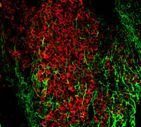
In the bone marrow, multiple myeloma cells are embedded in a niche defined by mesenchymal stromal cells, endothelial cells and immune cells. The non-hematopoietic stromal and endothelial cells provide survival and proliferation signals to the tumor cells are thought to be important determinants of the therapy responsiveness of multiple myeloma cells. Even though the functional importance of the non-hematopoietic niche is well established, the identity of the myeloma-supportive niche cells and the mechanistic underpinnings of this support are less well clarified.

Building on our experience with stromal cell biology in lymphoid organs we have set up an analyses pipeline to identify and characterize alterations in non-hematopoietic niche cells in multiple myeloma patients. We use single cell sequencing, multicolor flow cytometry and 3D culturing to identify and mechanistically interrogate primary stromal cells from myeloma patients. Experimental models of multiple myeloma allow us to unravel the importance of niche-derived signals for multiple myeloma disease progression and therapy response.
Circulating Tumor Cells
Careful analyses of peripheral blood of multiple myeloma patients has revealed the presence of tumor cells in circulation. The biological significance of these cells remains unclear, and could either reflect non-specific spill from an initial bone marrow location or active dissemination to secondary bone marrow sites. Understanding the composition and biological mechanisms of circulating myeloma cells could allow for disease monitoring and tumor cell analyses using peripheral blood, without the need for invasive bone marrow sampling.